Sonny James
IACT Certified Clinical Thermographic Technician
Managing Director, Thermal Diagnostics Limited ~ Medical Division ~
15 Robertson Street, Les Efforts EastSan Fernando, Trinidad & Tobago,
West Indies
868-653-9343 / 868-657-6572www.infraredclinic.com
Abstract
Digital Medical Thermal Imaging has been around for several decades and has proven itself time and time again to patients and doctors. The benefits of such imaging to both the physician and patient are invaluable when a diagnosis via conventional structural imaging and standard physical examinations are inconclusive or do not provide relief to the patient.
This paper will discuss the benefits of Medical Thermography for both men and women from head to toe. It will also discuss women's breast imaging and why all women should get imaged with thermography. Several intriguing case studies that the author came across at his Medical Thermography imaging center over the past year will be presented as well.
Introduction
At the IR/INFO 2010 conference, I presented a paper titled “How and Why I Got Into Medical Thermography”. This paper was a precursor to the information contained here. For those interested in my previous paper, you may view it at the IR/INFO website.
Although Medical Thermography has been around for several decades, it has not received the attention and credit it deserves from the medical establishment. However, we are seeing more patients becoming aware of this amazing technology. Usually it is the patient that ends up informing their physician about thermography.
Discussion
Unlike other imaging technologies such as ultrasound, mammography (radiography) and Magnetic Resonance Imaging (MRI), thermography is really in a class by itself. All of these other technologies look at the human body in almost the same dimension; that is, the structure or anatomy of the human body. Thermography does not look at the human body in this way. Thermography is a test of function or physiology.
Because Medical Thermography is a test of function rather than structure, it is capable of alerting you and your doctor of problems that are being missed or even misdiagnosed. Thermography sees what radiography, ultrasound and MRI cannot (and vice versa). So, right away we see that all of these technologies add to one another and do not replace each other.
For example, many patients who suffer from pain issues would usually get an x-ray or MRI. These technologies will look inside the body and give the physician an image of the internal structure or make-up of the body. Bones, organs, tumors and even blood vessels can be seen, but the dilemma that doctors meet is when nothing abnormal is seen with the internal structure of the human body. Everything is where it is supposed to be and no lumps or tumors are noticed. This is very frustrating to the doctor because the job of diagnosis tends to now fall mainly with the patient’s symptoms. Sometimes this is not enough and usually leads to an educated deductive reasoning (guess) by the doctor so that some kind of treatment (even if it is only for the symptoms) can be administered. In the meantime, the patient is still in pain and the root cause of the problem is not found.
Clearly we see that the doctor is not getting all the relevant information and only has one piece of the pie. This is where Medical Thermography comes in. Thermography shows the patient and doctor what is being missed. It adds an extra piece to the pie. Thermography shows how the body is functioning and how it is being physiologically affected by the patient’s problem. Many ailments and medical problems start off as a physiological change before structure is affected. Problems associated with inflammation, nerve root irritation, trigger points, muscular spasms, hormone imbalance and circulatory issues can effectively be seen with Thermography. Thermography can lead the doctor in the right direction to properly diagnose and treat the patient.
Our Medical Division
We have been providing Medical Thermographic Imaging for several years and have come across many interesting cases. Our business is comprised of about 50% breast imaging and 50% body imaging. We are very small with a staff of only two technicians, but still keep pretty busy.
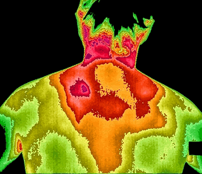
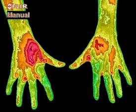
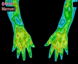
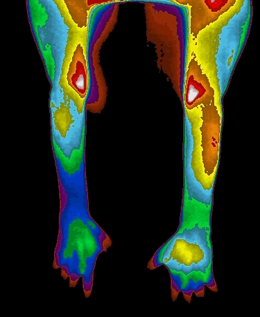
The following are some thermographic images of just a few cases during the last year: From head to toe…
Dental Issues
|
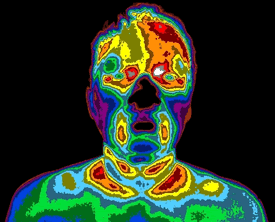
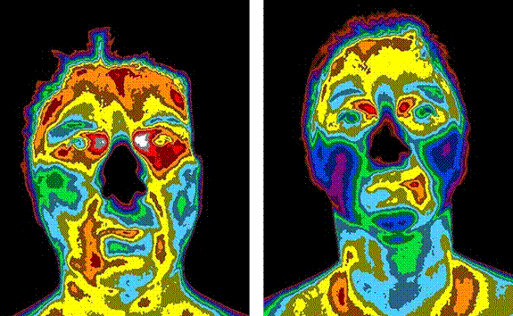
Patient with asymptomatic dental irritation seen over the right mandibular area (indicated by the abnormal heating on the left side of the image). |
Patient with asymptomatic dental irritation seen over the left mandibular area (indicated by the abnormal heating on the right side of the image). |
Stroke Screening
Patient came in for body imaging for a separate, unrelated problem. Thermogram of face shows the right side of the forehead and eye (left side on image) abnormally cool.
This thermal signature is suggestive of either arterial malformation or narrowing or hardening of the arteries. This is a sign of a possible impending stroke.
Thyroid Function
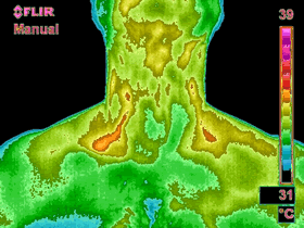
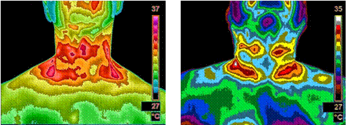
Image shows normal thyroid function
|
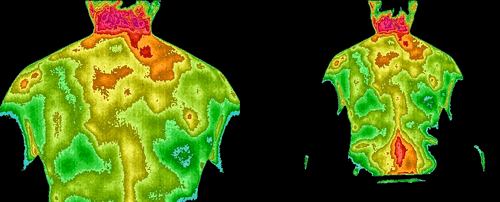
Images show signs of thyroid dysfunction and warrant thyroid work-ups. |
Breast Imaging and Breast Cancer Screening
Medical Thermography has excelled in breast cancer screening. Many times, Breast Thermography shows the earliest warning signs that a problem exists. All women 18 years and up should get at least one Breast Thermography done as it is documented that it is one of the most sensitive imaging modalities for breast disease and cancer screening. The following images all show evidence of abnormalities that were not picked up by mammograms or ultrasound.
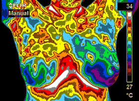
40 Year-old Patient:
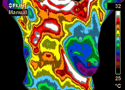
Right Breast (left side on image) graded as abnormal (TH4).
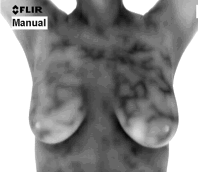
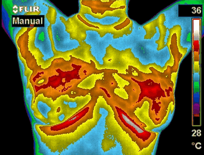
Patient with a unique vascular pattern suggestive of the effects of hormones on the breasts.
It is common knowledge that one of the contributing factors for higher risk of breast cancer is a hormone imbalance such as estrogen dominance. A prolonged or lifetime exposure to unopposed estrogen puts all women at higher risk of developing breast cancer.
Thermography can catch hormone imbalance issues early and can also be used to monitor treatment. Catch it early, fix it early and possibly prevent breast cancer. This is true early detection and prevention unlike any other imaging modality.


