Sonny James
IACT Certified Clinical Thermographic Technician
Managing Director
Thermal Diagnostics Limited – Medical Division
15 Robertson Street, Les Efforts East San Fernando, Trinidad & Tobago, West Indies868-653-9343/868-657-6572 www.infraredclinic.comAbstract
Thermography is one of the most widely-applied nondestructive test technologies in the world. Like many other NDT technologies, thermography has also made its way into the medical field. Thermography can be used to help detect a wide range of conditions including soft tissue injuries, inflammations, circulatory problems, and breast cancer.
This paper discusses how Medical Thermography differs from other diagnostic modalities along with challenges it has had and currently has with peer acceptance. This paper will also discuss the journey the author went through to introduce Medical Digital Infrared Imaging within his region.
Introduction
Thermography is one of the most diversified nondestructive testing and condition monitoring technologies. Thermography is used in almost every aspect of an industrial and commercial site. From the standard electrical inspection to the very complex furnace tube diagnostic. Thermography is now one of the most publicly known nondestructive testing technologies in the world. There are more video clips on YouTube of thermography than any other NDT application.
As like most other NDT methods and technologies, thermography has also made its way into the medical field. In medicine, Medical Thermography is commonly referred to as Digital Infrared Imaging. However, it has not fully received the medical industry’s acceptance or recognition, as one would think it has.
What Makes Medical Digital Infrared Imaging Different
Unlike other imaging technologies out there such as ultrasound, mammography and Magnetic Resonance Imaging (MRI), thermography is really in a class by itself. All of these other technologies look at the human body in almost the same dimension; that is, the structure or anatomy of the human body. Thermography does not look at the human body in this way. Thermography is a test of function or physiology. Therefore, it is sometimes harder to convince or quantify a function using thermography rather than an actual structural mass that can be measured and located with extreme precision using UT, MRI or x-ray scanning.
The Misinformation and Politics of Medical Digital Infrared Imaging
Thermography as a medical application has another major hurdle to overcome that will take some time to be fully accepted and embraced by mainstream medical professionals and associations. Qualified and experienced industrial thermographers can relate and understand how thermography has received a bad name mainly because it is sold as and considered an easy NDT application and that anyone with an imager can carry out an inspection. This is not true and has dramatically hindered the proper progress and promotion of this fascinating technology. The exact same thing has happened in the medical field. In the early stages of medical testing, anyone and everyone with a thermal imager was performing Medical Digital Infrared Imaging. This led to outrageous claims and findings that were easily challenged and discredited by medical professionals. There were no standards or protocols for equipment and no requirements for the qualification and certification of medical thermologists. Interpretation was also very subjective and no acceptable criteria were established or put in place. These were the perfect ingredients for the discrediting and failure of thermography within the medical field.
If only medicine and medical professionals were as proactive and forgiving to technology as the NDT community. Again, experienced NDT professionals know that every technology has its advantages and limitations. They also know that only properly trained and qualified inspectors are capable of interpreting and evaluating results from any NDT method. Even though a viable and useful technology is found to be ineffective at first, designers and NDT professionals work together to perfect it. This is because they know that it will eventually help all NDT inspectors and engineers with plant and equipment diagnostics, thus saving money and lives. In addition, there is much profit to be made with a new and effective technology.
It is not so in the medical industry. Stakes are much higher than just plant equipment. They are dealing with human lives and the diagnostics of human health. Small mistakes can lead to very huge lawsuits and even the discrediting of very skilled doctors and medical professionals. That is one main reason why progress moves so very slowly in medicine. There is also the fear of having a new technology being used as a replacement to another technology that has been used as the standard and mainstream for certain diagnostics. The fear of profit loss for certain equipment makers, medical associations, and doctors is a great deterrent to progress. Many doctors and medical professionals are not as educated about diagnostic and testing technologies as are NDT professionals. NDT professionals know that one NDT technology does not replace another; all technologies and methods compliment each other in the ultimate goal of problem diagnostics and root cause analyses. Something as old as liquid penetrant testing has not been replaced by eddy current or magnetic particle testing.
Progress is Truly Inevitable and Unstoppable
Now there is hope and a bright future for Medical Digital Infrared Imaging. A certain few doctors and medical professionals out there have seen and are convinced of the medical benefits of thermography and have been working hard to have it accepted by their peers. With advancements in infrared detector technology and the establishment of standards, protocols and practices, Medical Digital Infrared Imaging is making a strong comeback. What is also a strong driving force is that patients are becoming more educated and aware of Medical Digital Infrared Imaging and its benefits because of the open information on the internet and in the media. The patients are now educating and raising the awareness of their doctors. Just like in NDT, “industry” (patients) are demanding better healthcare and preventive medical care and diagnostics.
This is great news, but we should be cautious, hold restraint, and not have the “full steam ahead” mentality that initially led to problems for Medical Digital Infrared Imaging. We should continue to research and understand the future capabilities of this technology while at the same time using thermography on patients with applications that we all know work and have already been adequately studied. There are uses for Medical Digital Infrared Imaging right now and it is actually saving lives as we speak and enabling people to live a better quality of life.
The Ten Year Journey – Our Story
I have been involved in infrared thermography and NDT for over seventeen years. My first encounter with Medical Digital Infrared Imaging was ten years ago when I had a friend whose car mechanic got his finger punctured in a work related injury. For weeks, this person was going to his family doctor with no signs of relief or healing; in fact, the problem seemed to be getting worse with swelling and pain. Therefore, we decided to look at it with my thermal imager that I use for electrical inspections. What I saw was a very cold digit that appeared to be lacking proper blood circulation at the area of the injury. I am in no way qualified to interpret or evaluate medical thermograms but I did tell him to see his doctor with these images and that he should do something to improve the circulation in his finger.
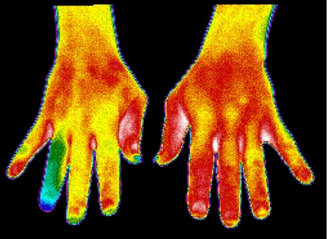
Insufficient blood flow to patient’s right hand ring finger
A few weeks later, I got a call from my friend who told me that his mechanic took my advice and has made a full recovery.
That was it! I was now bit by the Medical Thermography bug. So, I did some research and made some brochures and began going to almost every chiropractor and sports injury doctor I could find in my region. Every single doctor shot me down! They agreed that it was an interesting technology, but did not want to spend any money to invest in it for their patients or even just have me come and perform some scans for them to help with their diagnoses. They all told me that they have x-ray for that. Therefore, I ultimately gave up and returned to my circuit breakers and boilers.
Life was good, it was business as usual, and several years went by. However, I never forgot about how good thermography is at finding potential health problems. Early in 2008 I was doing more research on Medical Digital Infrared Imaging and discovered Dr. William C. Amalu. A chiropractor from San Francisco, California who has, together with other respected doctors, made positive advancements in Medical Digital Infrared Imaging, especially in the breast health application. I contacted him and inquired about the prospects of establishing a testing clinic in my country. To my surprise, Dr. Amalu’s answer was “yes”, as long as I follow established standards and protocols and be trained and certified as a medical thermographic technician. For obvious reasons, he also recommended that I get a female technician involved. So I spoke to my wife and she was willing to try it.
Being a skeptic myself and wanting to research everything before I gambled my money and good name, I still needed more unbiased information. So, I did some more research on Breast Thermography and Dr. Amalu. All looked great. Nevertheless, I was still a little hesitant, until one night when I decided to use my imager and look at my wife.
From what I saw on the internet and literature on what a “normal” breast thermogram should look like, I was very concerned about my wife’s images. I was seeing a lot of thermal activity that should not be there. I immediately contacted Dr. Amalu and sent him my images. He was worried enough with what he saw that he explained to me in detail what I needed to do to retake the images in the proper way that would enable proper interpretation. I had to follow protocol to allow proper interpretation.
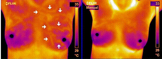
After following the detailed instructions, I took a series of images and sent them to Dr. Amalu for his interpretation. The results were her right breast was normal but her left breast was between normal and abnormal (also referred to as questionable) with signs of a hormonal problem. Dr. Amalu recommended that my wife see her doctor to have a follow-up mammogram (complementary test) and further tests to determine the cause of the hormonal signs.
Two months went by and after working with her doctor on corrective measures to her health, we decided to take another series of thermograms to see if there were any changes. The results were amazing. Activity in the left breast decreased significantly. Actual progress was achieved and seen. Left unaddressed and unnoticed, her condition could have developed to a more serious condition and possibly even breast cancer.
 |
|
|
Left breast graded as questionable |
After two months of corrective measures |
In January 2009, both my wife and I flew to San Francisco for training. Upon our return, we established a small test center and started aggressive advertising. I gave several radio interviews and made a TV commercial. Now armed with the right information, I contacted the right doctors and introduced what we were doing to help women. All the doctors were very interested and happy to have this technology finally in the region. We are now getting referrals from doctors and the public is slowly becoming aware of this life saving technology. Just recently we found out that health insurers in our region have decided to cover this test and most cover up to 80% of the cost. The future for Medical Digital Infrared Imaging in our region is looking bright, and more and more women are being helped in ways they thought they never would be.
So What is Medical Digital Infrared Imaging and How Does it Work?
Medical Digital Infrared Imaging is unlike any other medical screening diagnostic technology. As mentioned earlier, we are not looking at the structure or anatomy of the human body, but rather looking at the function. We cannot see “inside” the body. We are only seeing surface thermal or heat patterns that are being radiated naturally by the human body. In basic terms, when there is a physiological or metabolic change or problem in the human body, it manifests itself as a heat or lack of heat signature.
Let us take for example certain breast cancers. Cancer in simple terms is defective DNA and cells multiplying and growing. In order for these cancer cells to grow, they need nutrients. Their main source of nutrients comes from the blood. Therefore, what they do is awaken the dormant blood vessels within the breast or sometimes create a new blood supply to feed the cancer cells. The creation of the new blood vessels is termed “angiogenesis”. Humans are warm blooded and the blood vessels in the breasts are close to the surface of the breast. Therefore, the heat is transmitted to the surface of the breast and can be seen with the right thermal imager. If a problem in the human body causes a biochemical reaction (vascular and/or neurophysiologic) of sufficient magnitude, a thermovascular signal can manifest onto the surface of the body, then thermography can be used to help in diagnosis. Often times, thermography can find problems and abnormal health issues before other structural types of diagnostic technology can.
Think of it from a condition monitoring point of view. We as industrial thermographers know that in many cases, thermography can locate motor misalignment problems before vibration analysis and other technologies. That is because the first sign of trouble is manifested though heat – just like in the example for certain breast health issues.
And just like the analogy with the motor misalignment, we know that even though thermography can find signs of misalignment problem first, it cannot be used to quantify the misalignment or damage caused to the bearings. You will need vibration analysis and other technologies for that.
The same thing is done with Breast Thermography. We usually find issues before they can be found with other structural technologies. Therefore, the doctors will always recommend follow-ups with ultrasound, mammogram, or MRI to make sure that nothing structural is being missed. If nothing structural can be found, it is recommended that more frequent thermography follow-ups be done in order to monitor the condition of the thermal abnormality. In addition, the patient’s doctor may try different treatments to see if the abnormal condition can be improved. Only the doctor and patient can decide what should be done to a patient’s health. Perhaps it may be close monitoring until the abnormality has grown to the size that can now be identified with structural imaging technologies. Once the doctors can find it and size it, they already know that it is affecting the patient’s function, so now they can diagnose and attack the problem. This is true early detection and the difference between life and death.
Remember that thermography is not a stand-alone technology. Just like any other NDT method, it requires the addition of other technologies for proper diagnosis and treatment. The same can be said for mammograms, ultrasound, and MRI. All these technologies are great, but alone they offer minimal sensitivity and accuracy. Combined, true predictive and preventative medical care can be achieved.
It is documented that in breast cancer detection, mammograms alone are at most 80% sensitive and specific; thermography alone is ~90% sensitive and specific. However, combining Mammography + Thermography + Clinical Exams gives a greater than 95% chance of detecting early stage cancers.
The following is a list of what Medical Digital Infrared Imaging can be used to help detect:
|
|
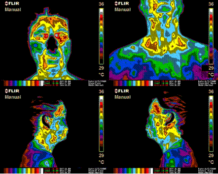
Female patient with multiple chronic pain conditions
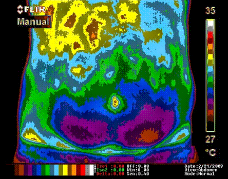
Male patient with upper right quadrant abdominal problems
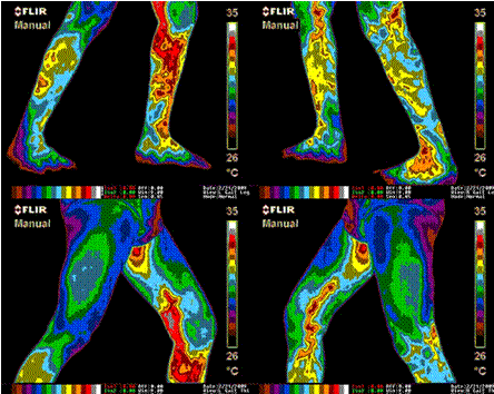
Male patient with severe varicosities (varicose veins)
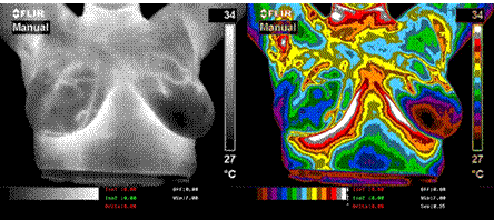
Right breast classified as abnormal (TH-4)
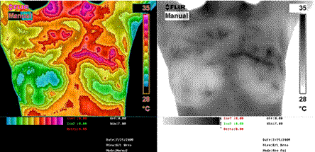
24-year-old patient. Left breast classified as abnormal (TH-4)
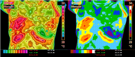
Right breast classified as very abnormal (TH-5)
Conclusion
Discussions can go on forever as to the viability of thermography in medical applications. However, I have personally seen the benefits and results, not on just my wife but on the patients we scan every week. I never get into any venture unless I truly believe that it works and can actually help people and companies. I decided to diversify into Medical Digital Infrared Imaging because I believe and know that this can help people throughout my region live a better and healthier quality of life.
Thermography has been approved by the FDA since 1982 as an addition to other screening technologies and has gone through many peer studies. I recommend that all women over 18 regardless of health history or how they are feeling get at least a baseline Breast Thermography scan done.
If we monitor and trend our health as we do our plant equipment, then less
of us will need to be repaired, overhauled or even worse, replaced!
Visit out Sponsors:
Electrophysics


